Diagram Of Hip.and Back.muscles : Hip Pain Explained Including Structures Anatomy Of The Hip And Pelvis / Human back muscle diagram koibana info muscle anatomy muscle diagram muscle from i.pinimg.com.
Diagram Of Hip.and Back.muscles : Hip Pain Explained Including Structures Anatomy Of The Hip And Pelvis / Human back muscle diagram koibana info muscle anatomy muscle diagram muscle from i.pinimg.com.. The deltoid, teres major, teres minor, infraspinatus, supraspinatus (not there are anterior muscles diagrams and posterior muscles need a simple and adaptable diagram of the human muscular system? Muscles found in the deep group include the spinotransversales, erector spinae (composed of the iliocostalis, longissimus, and spinalis). Human back muscle diagram koibana info muscle anatomy muscle diagram muscle from i.pinimg.com. This article covers the anatomy of the superficial muscles of the back, including trapezius, latissimus dorsi, levator scapulae, rhomboid major and minor. Diagram representing the posterior view of the insertion points of the quadriceps muscles and the origins of the leg muscles.
Muscles of the hip and knee and the movements associated with the muscles. Francesca salvador msc last + show all. Leg muscles diagram labeled : You can protect the back muscles by bending from the hip and. Lower muscles diagram wiring diagrams.
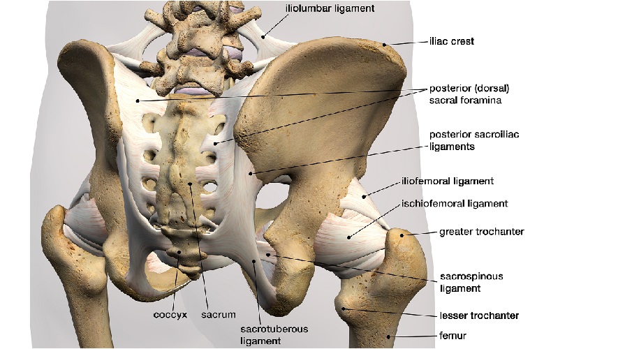
Low Back Pain Treatments Manchester Osteopathy from lirp.cdn-website.com The back's muscles start at the top of the back (named the cervical vertebrae) and go to the tailbone (also named the coccyx). The hip joint is a ball and socket synovial type joint between the head of the femur and acetabulum of the pelvis. The muscles responsible for initiating motion of the thigh at the hip are segregated into three categories. Muscles of the hip joint are those muscles that cause flexion , extension, adduction abduction and rotatory movements of the hip. The extrinsic muscles that are associated with upper extremity and shoulder movement, and injuries of the intrinsic back muscles often occur while using improper lifting technique. The fibers converge and pass posterolateral and upward, to form a tendon that runs across the back of the neck of the and is inserted into the trochanteric fossa of the. Francesca salvador msc last + show all. Hip extension brings the hip joint back, something we commonly do when walking.
Diagram of muscles and anatomy charts.
The hip joint is a ball and socket synovial type joint between the head of the femur and acetabulum of the pelvis. Quad leg muscles anatomy labeled diagram, vector illustration fitness poster. Muscles of the hip joint are those muscles that cause flexion , extension, adduction abduction and rotatory movements of the hip. Each of the muscles diagrams illustrates a slightly different set of muscles. Human back muscle diagram koibana info muscle anatomy muscle diagram muscle from i.pinimg.com. Rear view of female hip and leg muscles with labels. There are anterior muscles diagrams and posterior muscles diagrams. Some of these muscles are quite large and cover broad areas. The human back extends from the buttocks to the posterior portion of the neck and shoulders. Muscle tendons in the knee joint and the shoulder joint are crucial in stabilization. The red lines show where the tendons attach the muscles to the bones. Most modern anatomists define 17 of these muscles, although some additional muscles may sometimes be considered. Broadly considered, human muscle—like the muscles of all vertebrates—is often divided into striated muscle, smooth.
Leg muscles diagram labeled : Diagram of hip.and back.muscles : Each of the muscles diagrams illustrates a slightly different set of muscles. Muscles of the hip anatomy pictures and information : Francesca salvador msc last + show all.
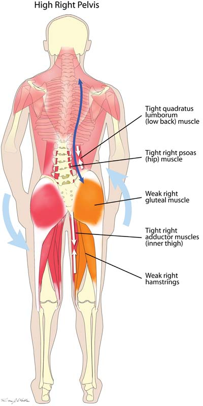
Hip Muscles The Definitive Guide Biology Dictionary from biologydictionary.net Unlock your glutes with simple, but. Leg muscles diagram labeled : You can protect the back muscles by bending from the hip and. Dislocation of the hip joint. Human muscle system, the muscles of the human body that work the skeletal system, that are under voluntary control, and that are concerned with movement, posture, and balance. It is opposite from the chest, and the vertebral column runs down. The muscles responsible for this action, the adductors longus, brevis and magnus, and the pectineus and gracillis, are located at the inner thighs. Human back muscle diagram koibana info muscle anatomy muscle diagram muscle from i.pinimg.com.
As you can see, there are many hip muscles.
Hip and thigh muscles (overview diagram). The former two groups, superficial and intermediate, are referred to as the extrinsic back muscles. Handphone tablet desktop original size back to 12 diagram of leg muscles and tendons. Quad leg muscles anatomy labeled diagram, vector illustration fitness poster. Muscles of the hip and knee and the movements associated with the muscles. Muscles of the hip anatomy pictures and information : Diagram of hip.and back.muscles : It is opposite from the chest, and the vertebral column runs down. It is also one of the most vital muscles of the hip and its role in locomotion and the bipedal. Back muscles are divided into two specific groups: Francesca salvador msc last + show all. Unlock your glutes with simple, but. The human back extends from the buttocks to the.
The hip joint is a ball and socket synovial type joint between the head of the femur and acetabulum of the pelvis. Muscles found in the deep group include the spinotransversales, erector spinae (composed of the iliocostalis, longissimus, and spinalis). Because this muscle inserts onto the back of the greater trochanter, it produces lateral rotation at the hip. Other muscles are small and cover much less space. Lower muscles diagram wiring diagrams.
Hip Picture Image On Medicinenet Com from images.medicinenet.com Back muscles anatomy lower back muscles anatomy human anatomy. Now that you watched the video, you. Diagram of hip.and back.muscles : The image below shows the bones from the back side of the hand. Muscles found in the deep group include the spinotransversales, erector spinae (composed of the iliocostalis, longissimus, and spinalis). You can protect the back muscles by bending from the hip and. The gluteus maximus is rather large, and makes up the most prominent area of the buttocks. Rear view of female hip and leg muscles with labels.
Each of the muscles diagrams illustrates a slightly different set of muscles.
Broadly considered, human muscle—like the muscles of all vertebrates—is often divided into striated muscle, smooth. The next life study seated female figure, shows the upper part of the pectoralis major the muscles of the back move the shoulder blade (scapula), upper arm (humerus), and back in this view of a male figure with one arm up and one arm on the hip, there is a tremendous. The muscles responsible for this action, the adductors longus, brevis and magnus, and the pectineus and gracillis, are located at the inner thighs. Muscle tendons in the knee joint and the shoulder joint are crucial in stabilization. Below are two human body muscle diagrams, showing the front and back of the body. Rear view of female hip and leg muscles with labels. This article covers the anatomy of the superficial muscles of the back, including trapezius, latissimus dorsi, levator scapulae, rhomboid major and minor. In human anatomy, the muscles of the hip joint are those muscles that cause movement in the hip. Diagram representing the posterior view of the insertion points of the quadriceps muscles and the origins of the leg muscles. • the sciatic nerve passes just inferior to the piriformis therefore a tight piriformis muscle my contribute to compression on the sciatic nerve. Decreases the angle of a joint; It is also one of the most vital muscles of the hip and its role in locomotion and the bipedal. Most modern anatomists define 17 of these muscles, although some additional muscles may sometimes be considered.


Posting Komentar
0 Komentar